Degenerative Myelopathy
(degenerative myelopathy, DM), formerly called “chronic degenerative radiculomyelopathy”, is a progressive, neurodegenerative disease of dogs that primarily affects the white matter of the spinal cord; during the course of the disease the nerve pathways of the spinal cord's white matter are gradually damaged, especially the nerve pathways that coordinate the movement of the hind body. The clinical and pathological features of degenerative myelopathy show similarities in many respects to the human disease amyotrophic lateral sclerosis (ALS), which is why the disease is of particular importance from the point of view of veterinary neurology and comparative medicine as well.
Degenerative myelopathy is a progressive and gradually worsening disease that is observed in adult/older dogs (generally ≥ five years old); it first affects the hind limbs and involves upper motor neuron ataxia and gradual paralysis, all the way to paralysis of the whole body. This pathology was first described in 1973 by the veterinarian Damon R. Averill Jr. in a German Shepherd dog, in which degeneration of unknown origin of the spinal cord, especially the white matter, was observed.
The research group of the University of Missouri (Zeng et al., 2014) analysed the genetic data of more than 33,747 dogs from 222 dog breeds. The main mutation associated with degenerative myelopathy was detected in 124 different breeds. The authors emphasised that the real number may be even higher than this, because for many breeds only a few individuals were examined. As the study puts it: the SOD1 mutation associated with the disease was present in 56% of the breeds examined, that is, in 124 different breeds.
The full original list containing 124 breeds is rather long, therefore the breeds most frequently mentioned in the literature, and the ones most affected with respect to the mutation, are the following:
High-risk breeds
In these breeds the SOD1 mutation is relatively common, and clinical cases of degenerative myelopathy have also been documented:
- German Shepherd
- Pembroke Welsh Corgi
- Boxer
- Chesapeake Bay Retriever
- Bernese Mountain Dog
- Rhodesian Ridgeback
- Cardigan Welsh Corgi
- Wire Fox Terrier
- Hovawart
The breeds studied so far in connection with the disease are available in a separate document.
A pathological approach
As regards localisation, the pathology affects the lateral and ventral funiculi of the spinal cord's white matter in the thoracolumbar segment (from T3 to L3). The distribution of the lesions varies from breed to breed: e.g. in German Shepherd dogs the lesions appear in a patchy, bilateral and asymmetric manner, whereas in Pembroke Welsh Corgi dogs they appear continuously along the length, within more clearly defined areas.
The aetiopathogenesis of this pathology (the triggering cause of the disease and the process of its development) is not clear, and over the years numerous hypothesised triggering causes have been considered: metabolic, nutritional, vascular, immune-mediated and/or vitamin E deficiency, but currently the most credible cause is thought to be genetic.
In 2006 the neurologist Dr. Roger Clemmons and his colleagues hypothesised a genetic connection between human multiple sclerosis (MS) and canine degenerative myelopathy. In the course of their work, Clemmons and colleagues carried out a multitude of genetic examinations, during which they observed in German Shepherd dogs suffering from degenerative myelopathy a gene mutation similar to human multiple sclerosis.
After this, in 2009 Dr. Toshio Awano identified a mutation affecting a single nucleotide in a specific region of chromosome 31, which contains the gene coding for superoxide dismutase (SOD1), one of the most frequently represented proteins in the central nervous system, whose specific function is the removal of free radicals. (scientifically: In the affected dogs, a substitution of guanine (G) for adenine (A) was observed at position 118 of exon 2 (c.118G>A), which led to a missense mutation of glutamic acid to lysine at position 40 (E40K) = SOD1:c.118G>A (E40K).)
In the development of the disease, therefore, researchers attribute a significant role to the mutation of the superoxide dismutase-1 (SOD1) gene, which plays an important role in the protection of nerve cells against oxidative stress. As a result of the faulty functioning of the gene, damage to the nerve tissue may increase, which in the long term leads to the degeneration of the nerve cell's axon and of the myelin sheaths.
The role of the spinal cord and the nerve pathways in degenerative myelopathy
From a pathological point of view, during degenerative myelopathy a progressive damage of the ascending and descending tracts of the spinal cord can be observed. The process begins primarily in the thoracolumbar (T3–L3) spinal segment, and over time may also spread in the cranial and caudal directions. During the disease, damage to the myelin sheath (demyelination), axonal degeneration and gliosis (scar formation in the nervous system) develop, as a result of which nerve conduction gradually deteriorates. The damage primarily affects the upper motor neuron pathways, which explains the development of spastic paresis (increased muscle tone in the limbs), hyperreflexia and ataxia (a disorder of movement coordination).
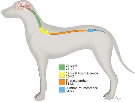
To understand degenerative myelopathy, it is important to briefly get to know the functioning of the dog's nervous system, especially the structure of the spinal cord and the nerve cells. The essence of the disease is that the nerve pathways in the spinal cord (mainly in the white matter) are gradually damaged, and because of this the communication between the brain and the limbs is interrupted.
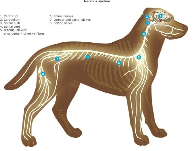
The nerve cell (neuron) is the fundamental structural and functional unit of the nervous system. In the dog's nervous system it works the same way as in other mammals: it transmits information in the form of electrical and chemical signals through the long processes (axons) between the brain, the spinal cord and the various parts of the body.
The structure of a nerve cell
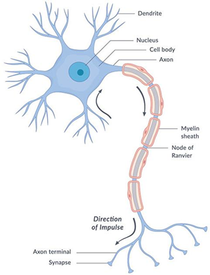
- Dendrites: short processes that receive signals from other nerve cells.
- Cell body (soma): the central part of the nerve cell, containing the nucleus.
- Axon: a long process that transmits the electrical impulse.
- Myelin sheath: the insulating layer surrounding the axon, which speeds up nerve conduction.
- Nodes of Ranvier: the small sections between the myelin sheath, where the impulse travels on “in leaps”.
- Axon terminals/synapses: these pass the signal on to other nerve cells or muscles.
How does the dog's nerve cell work?
- The sense organs generate a nerve impulse.
- The dendrites of the neuron receive the signal.
- The electrical impulse travels along the axon.
- The myelin sheath helps this impulse spread very quickly.
- At the end of the axon the signal is passed to another nerve cell or muscle cell.
What does the neuron mean in the dog's movement?
- running,
- balancing,
- the reflexes,
- the quick reaction to commands.
The impulse starting from the dog's brain travels along the axon, and the myelin sheath speeds up this process, so that the muscles are able to react very quickly.
During degenerative myelopathy, primarily the axons and the myelin sheaths are damaged.
The role of the axon
The axon is the long process of the nerve cell that transmits the nerve instructions starting from the brain, carrying electrical impulses towards the muscles. These impulses make possible:
- walking, running,
- balance,
- coordinated movement, tail wagging,
- as well as the functioning of the reflexes, reacting to a command.
The motor nerve cells of the brain send signals towards the muscles. These signals travel through the axons. The longer the distance (for example from the spinal cord to the hind legs), the more important the efficient functioning of the axon is.
When the axon is damaged, the impulse does not reach the muscles properly. This is why the following appear:
- the uncertain gait,
- the stumbling,
- the weakness of the hind legs,
- and later paralysis.
The role of the myelin sheath
The myelin sheath is the outer covering surrounding the axon, a fatty insulating layer around the axon. Its function is similar to the plastic insulation of electric cables.
Its task:
- protects the axon,
- speeds up nerve conduction,
- ensures precise nerve communication,
- saves energy for the nerve cell.
The myelin sheath makes possible so-called “saltatory conduction”, when the electrical signal does not travel continuously along the axon but “jumps” from section to section. Because of this the dog:
- can react faster,
- coordinates its movement more precisely,
- can stay balanced while running or jumping.
During degenerative myelopathy the myelin sheath gradually breaks down (demyelination). Because of this:
- the impulse travels more slowly, becomes imprecise,
- the nerve signals become distorted,
- and finally the axon may degenerate completely.
This explains why the symptoms gradually worsen.
If the axon or the myelin sheath is damaged:
- movement may slow down,
- the gait may become uncertain,
- weakness or paralysis may develop.
This may occur:
- in a spinal injury,
- in nerve inflammation,
- or in certain nervous-system diseases (see DM).
How does the SOD1 gene affect the functioning of the myelin sheath and the axon?
The SOD1 gene plays an important role in the protection of cells against oxidative damage. In dogs, certain SOD1 gene mutations are associated with degenerative myelopathy.
The role of the SOD1 gene: The SOD1 gene codes for the enzyme superoxide dismutase 1. This enzyme:
- neutralises the harmful free radicals,
- protects the nerve cells from oxidative stress,
- supports the long-term functioning of the nerve cells.
How does the mutation of the gene damage the axon and the myelin sheath?
The mutated SOD1 gene protein may work faultily and gradually damages the nervous system:
Axon damage - The axons gradually degenerate:
- nerve conduction deteriorates,
- the communication between the brain and the muscles slows down,
- the coordination of the hind limbs weakens.
Damage to the myelin sheath - Because of the breakdown of the myelin sheath:
- the nerve signals travel more slowly,
- the loss of impulse increases,
- an uncertain gait and muscle weakness develop.
What symptoms appear during the damage?
- weakness of the hind legs,
- dragging of the claws of the hind legs,
- loss of coordination,
- later paralysis.
Where does the damage occur?
The most significant lesions develop in the white matter of the spinal cord.
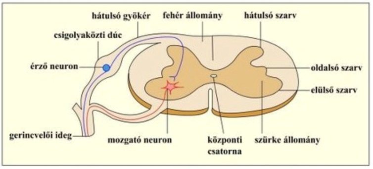
The spinal cord consists of two main parts:
- Grey matter - This is where the cell bodies of the nerve cells are located.
- White matter - This is where the nerve pathways run, that is, the axons covered with the myelin sheath. During degenerative myelopathy, mainly the white matter is affected.
Why are the hind legs affected first?
The disease begins in the thoracolumbar (T3–L3) spinal segment, which contains the nerve pathways that innervate the hind limbs.
This is why the following appear first:
- the coordination disorders of the hind limb,
- the ataxia,
- the dragging of the claws,
- the weakness of the hind body.
Later the damage progresses to other segments of the spinal cord as well, so the functioning of the front limbs and the respiratory muscles may also become affected.
What happens to movement?
Normally:
- The brain sends a movement command.
- The impulse travels through the spinal cord.
- The axons transmit the signal.
- The muscles contract.
In the case of degenerative myelopathy:
- the myelin sheath is damaged,
- the axons die,
- the impulse is interrupted,
- the muscles do not receive a proper signal.
This causes the gradual paralysis.
⚠️ Why is it important to understand this?
Many owners think at first that the dog:
- “is just getting old”,
- has joint pain,
- or is struggling with a hip problem.
Degenerative myelopathy, however, is a disease of nervous-system origin. The problem does not begin primarily in the muscles or the joints; rather, the nerve communication is damaged.
Understanding this can help:
- in early recognition,
- in choosing the appropriate rehabilitation,
- as well as in preparing owners for the progression of the disease.
The course of the disease
Degenerative myelopathy progresses slowly; at first the affected dogs show proprioceptive ataxia (a movement-coordination disorder caused by the loss of the perception of body position); in the initial stage these deficits often appear asymmetrically, later progressing with paralysis and then complete paralysis. In most dogs the clinical symptoms of the disease are consistent with upper motor neuron injury, with increased spinal reflexes, proprioceptive deficits and increased muscle tone; less frequently, decreased spinal reflexes and muscle tone have also been reported. One essential feature of the disease is that it does not cause pain. Most patients may lose the ability to walk within 6–9 months of the appearance of the symptoms. Following the paralysis of the hind limbs, the gradual loss of the spinal reflexes of the hind limbs, the gradual paralysis of the thoracic limbs, faecal and urinary incontinence, and signs of brainstem involvement such as dysphagia (difficulty swallowing) and, finally, breathing difficulties appear.
The clinical symptoms of degenerative myelopathy generally appear slowly and gradually. In the early stage, owners often observe mild coordination disorders, uncertainty of the hind limbs, stumbling, crossing of the hind legs, or abnormal wear of the claws. The symptoms initially resemble, in many cases, an orthopaedic problem, which is why recognising the disease is difficult. During progression, the proprioceptive deficit becomes more and more severe, then the weakness and partial paralysis of the hind limbs develop. In the later stages, muscle atrophy (muscle wasting), complete paraplegia (paralysis of the hind limbs), as well as disorders of urination and defecation may also appear. In advanced cases the process may spread to the front limbs and the respiratory muscles as well.
The symptoms of the disease gradually worsen; its course is divided somewhat differently in the various literature, but most sources distinguish 3–4 main stages.
The most commonly used clinical classification:
1. Early stage
In this phase the nerve damage taking place in the white matter of the spinal cord is still mild; however, the functioning of the nerve pathways innervating the hind limbs is already deteriorating. The symptoms are mild and easily confused with joint or orthopaedic problems or with ageing.
Characteristic symptoms:
Coordination disorder (ataxia); the dog's gait becomes uncertain:
- slightly swaying movement,
- crossing hind legs,
- unstable turning.
Dragging of the claws - One of the earliest symptoms:
- the dog does not lift its paw properly,
- the claws touch the ground,
- abnormal claw wear can be observed. In the next stage the claws wear down completely and bleed.
Proprioceptive deficit, during which the dog perceives the position of its limbs less well. For example:
- it corrects the position of the paw with a delay,
- “knuckling” – the dog steps on the upper part of the paw,
- it stands up with more difficulty,
- it trips over smaller obstacles.
Mild hind-limb weakness
- slower movement,
- reduced jumping ability,
- difficulty with stairs.
The dog shows no pain at all (unless it is accompanied by another disease that may involve pain).
2. Intermediate stage
In this phase the degeneration of the axons and the myelin sheath increases significantly, and together with it the weakness of the hind limbs. Therefore, in this stage the dog already:
- stands up with difficulty,
- falls frequently,
- is capable of shorter walks.
Spastic paraparesis, during which the hind legs:
- become stiff,
- their movement becomes uncoordinated,
- the tone of the muscles may increase.
Muscle wasting (atrophy). Because of the reduced nerve stimulation:
- the thigh muscles become thinner,
- muscle mass decreases.
Loss of balance
- falling over when turning,
- worsening instability on slippery surfaces.
Increased fatigue may occur; movement requires more and more energy.
In this phase many dogs already:
- require a harness, a wheelchair,
- a lifting sling,
- intensive rehabilitation help.
Many authors consider this the point at which the dog's quality of life begins to deteriorate faster.
3. Advanced stage
The spinal cord damage in this phase is already severe; the hind limbs are mostly paralysed.
Partial or complete paraplegia. The dog:
- is unable to walk on its own,
- drags its hind limbs (“seal-like” movement),
- or completely loses the use of them.
Severe proprioceptive disorder, during which the perception of the limbs' position is greatly impaired.
Urination and defecation problems because of the involvement of the spinal nerve pathways:
- urinary and faecal incontinence may develop,
- the control of the sphincter muscles decreases.
Risk of pressure sores because of immobility:
- the risk of pressure injuries increases.
Chronic muscle loss, that is, the musculature of the hind body breaks down significantly.
In this phase many dogs:
- use a wheelchair, a lifting harness,
- require intensive home care.
4. End stage
The neurodegeneration now affects not only the hind body, but may also spread to the entire spinal cord and partly to the brainstem.
Involvement of the front limbs because of the progression of the disease:
- the front legs may also weaken,
- the dog may become completely immobile.
Weakness of the respiratory muscles (in rarer cases):
- the functioning of the chest muscles may also deteriorate,
- swallowing and breathing difficulties appear,
- brain involvement, dysphonia, facial nerve paralysis
Complete dependence on care
- the dog is unable to change position on its own,
- requires continuous care.
Deterioration of quality of life. In this stage, one of the most important questions for the veterinarian and the owner is:
- the sustainability of quality of life,
- and the making of humane decisions.
Why is it important to know the stages?
Recognising the phases of the disease helps:
- in early diagnosis,
- in planning the rehabilitation as early as possible,
- in choosing the appropriate aids,
- as well as in the psychological preparation of owners.
Although the progression of degenerative myelopathy cannot be stopped, however:
- physiotherapy,
- hydrotherapy,
- regular mobilisation,
- and, with appropriate home care, some dogs can preserve their quality of life for a longer time.
The stages do not follow one another at the same pace and in the same form in every dog; the speed of progression may depend on:
- the genetic background,
- the age,
- the general state of health,
- as well as the application of rehabilitation treatments.
The progression of the disease is therefore different from individual to individual, but:
- from the first symptoms to severe immobility often 6–18 months pass,
- most dogs lose the ability to walk within 1 year of the appearance of the first symptoms,
- for the occurrence of complete paralysis, the final stage, the literature gives various figures, but generally 17–36 months (the 36 months being distinctly optimistic).
⚠️ Important note!
Degenerative myelopathy is in itself a painless, slowly progressing, gradually advancing disease, which is why it can largely be distinguished from, for example, IVDD, a herniated disc, a spinal tumour or arthrosis. The secondary problems (pressure sores, other injuries, joint diseases), however, can cause pain.
One of the biggest problems of early-stage degenerative myelopathy is that its symptoms are very similar to many other, much more common orthopaedic or neurological diseases. Because of this, DM is practically a “diagnosis of exclusion”: other diseases must first be ruled out.
What is specifically characteristic of early DM?
The most typical initial symptoms:
- gradually worsening hind-limb uncertainty
- paw dragging
- asymmetric wear of the claws of the hind legs
- coordination disorder (ataxia)
- the dog “does not know exactly where its leg is”
- no or minimal pain
- symptoms slowly worsen over months
The absence of pain is one of the most important distinguishing signs.
Diseases that can be confused with the symptoms of early degenerative myelopathy
1. IVDD, or within it a herniated disc
This is one of the most important differential diagnoses.
Similar symptoms
- hind leg weakness
- stumbling
- uncertain gait
- coordination disorder
What points more to IVDD
- pain is common
- back sensitivity
- sudden worsening
- crying, restlessness
- avoidance of jumping
- muscle spasm
What points more to DM
- slow progression over months
- absence of pain
It can usually be distinguished with an MRI.
2. Lumbosacral stenosis / cauda equina syndrome
Common mainly in large-breed dogs.
Similar symptoms
- hind-limb weakness
- difficulty standing up
- gluteal muscle weakness
- uncertain gait
Points more to this
- strong pain in the lower back
- raising the tail is painful
- avoidance of stairs
- difficulty sitting
- limping may alternate
In DM
- there is usually no lower-back pain
- a coordination problem dominates rather
3. Hip joint arthrosis or dysplasia
Very often confused with early DM.
Similar symptoms
- difficulty standing up
- “old-age” movement
- uncertain hind legs
- decreased activity
In the case of a joint problem, rather
- pain during movement
- stiffness after rest
- limping
- palpable joint pain
- improves with painkillers
In DM
- the dog usually does not show it as painful
- a neurological coordination fault is rather visible
An X-ray can help in establishing the diagnosis.
4. Fibrocartilaginous embolism (FCE)
A spinal cord “stroke”.
Similar symptoms
- hind leg weakness
- loss of coordination
Distinguishing signs
- very sudden onset
- often one-sided
- not progressive
- stabilises after a few days
DM
- worsens slowly
- bilateral and gradual
5. Polyneuropathy / peripheral neuropathy
A group of peripheral nerve diseases, nerve damage.
Similar symptoms
- weakness
- uncertain gait
- muscle wasting
In the case of polyneuropathy, rather
- the front legs are also affected early
- reflexes are decreased
- voice change
- a swallowing problem may appear earlier
The neurological examination is especially important here.
6. CCL rupture (cruciate ligament injury)
Sometimes early DM is interpreted as “limping”.
- one-sided limping
- pain
- avoidance of weight-bearing
- sudden onset
DM
- is rather of coordination origin
- not classic lameness
The challenges of diagnosing the disease
Establishing the diagnosis is a complex process, since currently there is no single examination method that on its own would confirm the disease with complete certainty in a living animal. Part of the diagnostic process is the detailed neurological examination, imaging diagnostics — especially MRI —, as well as the exclusion of the differential-diagnostic possibilities. It is important to distinguish degenerative myelopathy from other spinal or orthopaedic diseases, for example IVDD, a herniated disc, lumbosacral syndrome, tumorous lesions or hip joint degeneration. Genetic examinations can help in detecting the predisposition; however, a positive genetic result on its own does not mean the certain development of the disease. The correct diagnosis of degenerative myelopathy can be a challenge, since the definitive diagnosis can only be made by histopathological examination of the spinal cord, post mortem (after the dog's death).
The most important differences during the neurological examination:
| Examination sign | Early DM |
|---|---|
| Pain | usually none |
| Proprioception (position sense) | deteriorates |
| Spinal reflexes | normal or increased |
| Muscle strength | slightly decreased |
| Progression | slow, continuous |
The “proprioceptive deficit” is very characteristic of degenerative myelopathy; the dog corrects its folded-over paw with a delay.
What examinations are needed?
- neurological examination
- spinal MRI
- X-ray
- CT in certain cases
- blood test
- genetic test!!! for the mutation of the SOD1 gene. The result of the test can be: N/N – clear, A/N – carrier, A/A – at risk.
What can be “DM-suspicious” in the early stage?
- older age
- slower, gradual worsening
- no pain
- symmetrical hind-limb symptoms (although the symptom initially starts on only one hind limb)
- paw dragging, claw wear
- normal blood test results
- the MRI examination shows no significant compression
Closing words
According to current knowledge, degenerative myelopathy is not curable, and treatment is primarily aimed at slowing the progression and maintaining quality of life. Physiotherapy, hydrotherapy and the maintenance of regular movement can, in certain cases, contribute to preserving the functional state for a longer time. In addition, appropriate home care, the prevention of pressure sores, and the psychological support of owners are of particular importance. The long course of the disease places a significant emotional and physical burden on owners, which is why degenerative myelopathy is considered an important research area not only from a veterinary but also from an animal-welfare and psychological point of view. Research is being carried out intensively even today. The development of new genetic examinations, neuroprotective therapies and regenerative medicine may offer hope for future treatment possibilities. Early recognition, appropriate rehabilitation and the education of owners are currently among the most important elements of managing the disease.
References, scientific publications:
- Tomoyuki Awano, Gary S Johnson, Claire M Wade, Martin L Katz, Gayle C Johnson, Jeremy F Taylor, Michele Perloski, Tara Biagi, Izabella Baranowska, Sam Long, Philip A March, Natasha J Olby, G Diane Shelton, Shahnawaz Khan, Dennis P O'Brien, Kerstin Lindblad-Toh, Joan R Coates (2009): Genome-wide association analysis reveals a SOD1 mutation in canine degenerative myelopathy. https://pubmed.ncbi.nlm.nih.gov/19188595/
- Coates JR, Wininger FA.Vet Clin North Am Small Anim Pract. 2010 Sep; Canine degenerative myelopathy.
- Crisp MJ, Beckett J, Coates JR, Miller TM.Exp Neurol. 2013 Oct;248:1-9. doi: 10.1016/j.expneurol.2013.05.009. Epub 2013 May 23. Canine degenerative myelopathy: biochemical characterization of superoxide dismutase 1 in the first naturally occurring non-human amyotrophic lateral sclerosis model.
- Ivansson EL, Megquier K, Kozyrev SV, Murén E, Körberg IB, Swofford R, Koltookian M, Tonomura N, Zeng R, Kolicheski AL, Hansen L, Katz ML, Johnson GC, Johnson GS, Coates JR, Lindblad-Toh K.Proc Natl Acad Sci U S A. 2016 May 31. Variants within the SP110 nuclear body protein modify risk of canine degenerative myelopathy.
- Roger M. Clemmons DVM, PhDVeterinary Clinics of North America: Small Animal Practice 4, July 1992, https://www.sciencedirect.com/science/article/abs/pii/S0195561692500870
- Morgan BR, Coates JR, Johnson GC, Shelton GD, Katz ML.J Neurosci Res. 2014 Apr;92(4):531-41. doi: 10.1002/jnr.23332. Epub 2013 Dec 21. Characterization of thoracic motor and sensory neurons and spinal nerve roots in canine degenerative myelopathy, a potential disease model of amyotrophic lateral sclerosis.
- https://ivcjournal.com/integrative-management-degenerative-myelopathy/
- Merck Veterinary Manual – Parts of the Nervous System in Dogs https://www.merckvetmanual.com/dog-owners/brain-spinal-cord-and-nerve-disorders-of-dogs/the-nervous-system-of-dogs
- Merck Veterinary Manual – The Nervous System of Dogs https://www.merckvetmanual.com/nervous-system/myelin-disorders/myelin-disorders-in-animals
- Myelin Disorders in Animals - Nervous System https://www.msdvetmanual.com/nervous-system/myelin-disorders/myelin-disorders-in-animals
- UNESP – Canine spinal cord neuron and axon myelin sheath morphometry https://repositorio.unesp.br/entities/publication/27b55e6e-c955-4546-a2ce-a187d7679596
- PubMed – Myelin and oligodendrocyte development in the canine spinal cord https://pubmed.ncbi.nlm.nih.gov/26338416/
- https://www.petmd.com/dog/conditions/musculoskeletal/degenerative-myelopathy-dogs
- https://www.pdsa.org.uk/pet-help-and-advice/pet-health-hub/conditions/degenerative-myelopathy-dm-in-dogs
- https://www.veterinaryteachingacademy.com/blog/the-five-stages-of-degenerative-myelopathy
- https://dogmobilityproject.com/2025/12/05/understanding-the-stages-of-degenerative-myelopathy-in-dogs/
- https://shadeoutdm.com/helpful-items
Scientific databases:
- PubMed - One of the world's most important medical and biological databases.
- https://www.merckvetmanual.com/ - One of the world's most important medical and biological databases.
- https://ofa.org/ - One of the world's most important medical and biological databases.
- Google Scholar - An easier-to-use search engine for scientific publications.
- ScienceDirect - Contains detailed veterinary and neurological literature.
- Wiley Online Library - Very many veterinary journals can be found here.
- American College of Veterinary Internal Medicine (ACVIM) - Clinical recommendations and neurological professional materials.
- Merck Veterinary Manual - An excellent summary with professional descriptions.
🩺 The summary was professionally reviewed by
Dr. Ákos Pákozdy veterinarian, neurologist
(University of Veterinary Medicine Vienna, Austria)